State-of-the-Art Facilities
and Equipment
- HOME
- State-of-the-Art Facilities and Equipment
Facilities and Equipment
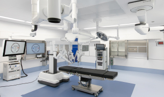
The Da Vinci Surgical System can deliver highly magnified, 3D high-definition views of the nerves and blood vessels of the surgical field. The Da Vinci System is designed to seamlessly translates the surgeon's hand movements into smaller, more precise movements by using tiny, wristed instruments that can bend and rotate far greater than the human hands. The Da Vinci System enables surgeons to perform even the most complex and delicate procedures through very small, incisions.
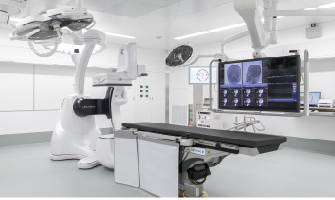
A hybrid operating room, which integrates imaging technology and surgical equipment to support a team of experts. This new suite also allows surgeons to perform procedures that are currently done open, in an endovascular (through the blood vessels) fashion instead. Many procedures are well-suited for hybrid ORs for the treatment of cardiovascular issues like coronary artery disease, valve replacement or repair.
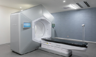
IMRT is the most advanced form of external beam radiation therapy available for cancer treatment today and precisely shaped beams of radiation are directed at a solid tumor. The precise control and flexibility of IMRT minimizes the amount of radiation going to surrounding healthy tissue. Less time on the table, which helps to minimize side effects. IMRT is used most often to treat prostate cancer or brain tumor.
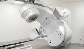
Linear accelerators are groundbreaking devices that are revolutionizing cancer treatment the LINAC can target and destroy cancerous cells in a precise area of a patientʼs body while minimalizing exposure to the radiation to the surrounding healthy tissue. Many types of cancer can be treated with this technology including, head and neck tumor, breast cancer, skin cancer, metastatic bone tumor.
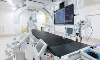
We have four angiography suites, and they can provide three-dimensional imaging during complex interventions with the aim of increasing the accuracy and safety of procedures. The Angiography suite is used for patients undergoing cardiology and vascular procedures including arrhythmia treatment (catheter ablation), endovascular treatment, cerebrovascular diseases and liver cancer.
Diagnostic Imaging
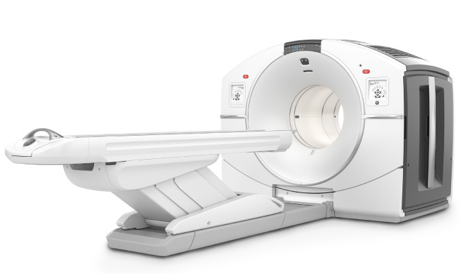
PET visualizes cancer and CT obtains position information. These have been combined to form one examination machine. Since the results of these two tests overlap, a more precise diagnostic image is possible, which is useful in the early detection of minute cancer.

The world's first PET scanner exclusive for the breast has been introduced. Since the breast is not pressed, the examination is painless. The women-friendly device can clearly visualize small, hard-to-find breast cancers.
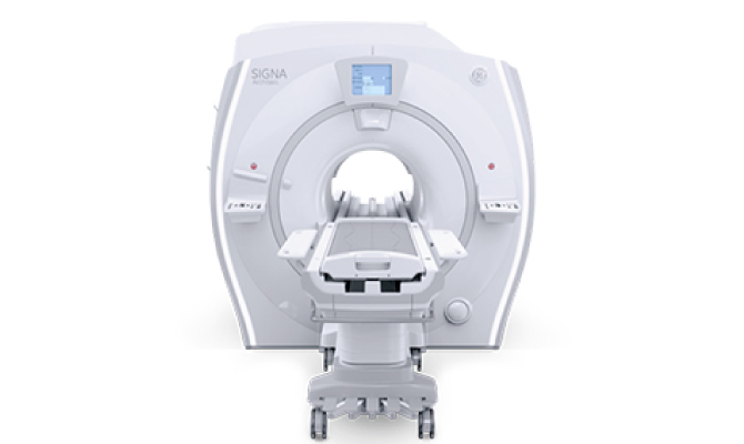
The equipment uses magnetism and radio waves, and inside the body can be visualized. Since there is no exposure to X-rays (radiation), a person can receive an examination with peace of mind. By visualizing various tissues, effective early detection of adult diseases is possible.
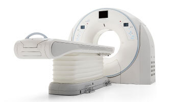
An image with a width up to 16 cm can be captured in one shot only in 0.35 seconds. It demonstrates ability in testing cardiac diseases such as angina pectoris, myocardial infarction, etc.
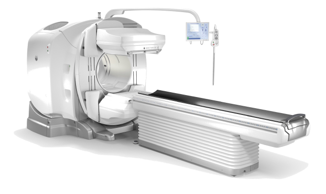
The equipment can combine SPECT with CT in order to diagnose various functions such as metabolism and blood flow. This is useful in the early detection of dementia as represented by Alzheimer's.
